
Ear Diagram Helix Human Anatomy diagram
Ear Anatomy, Diagram & Pictures | Body Maps Human body Head Ear Ear The ears are organs that provide two main functions — hearing and balance — that depend on specialized receptors called.
ear anatomy diagram labeled
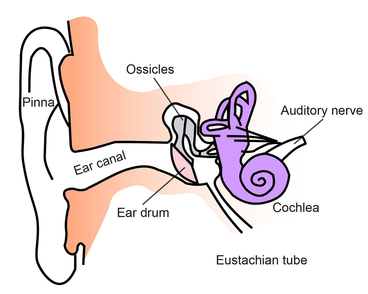
So as the air vibrates even the ear drum starts vibrating. Just like the skin of a drum. And as you can, the ear drum also separates the outer ear from the middle ear. This brings us to the middle ear. The middle ear consists of the three tiniest bones of the human body. And they're together the are called the ossicles. And they have pretty.
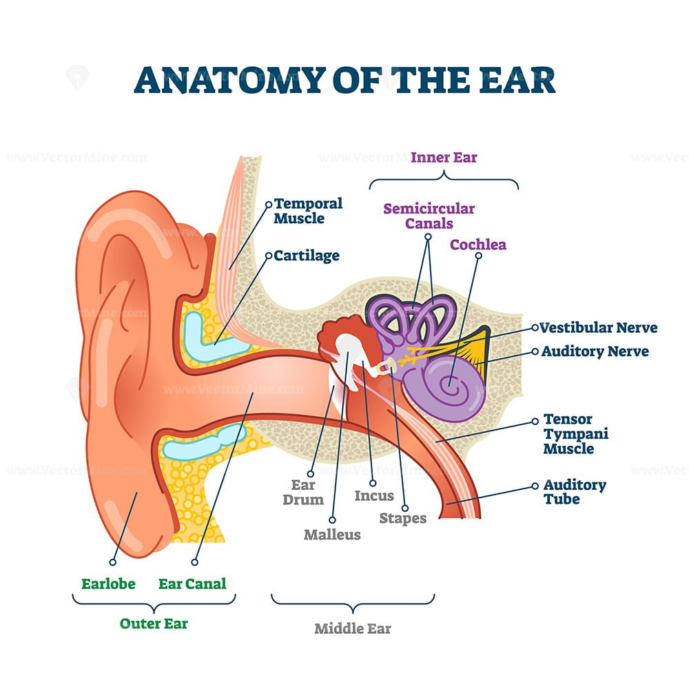
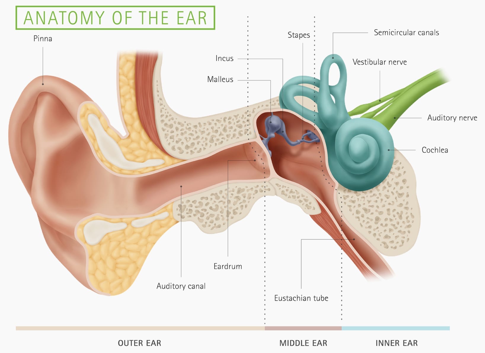
Anatomy of the ear, labeled health care vector illustration diagram VectorMine
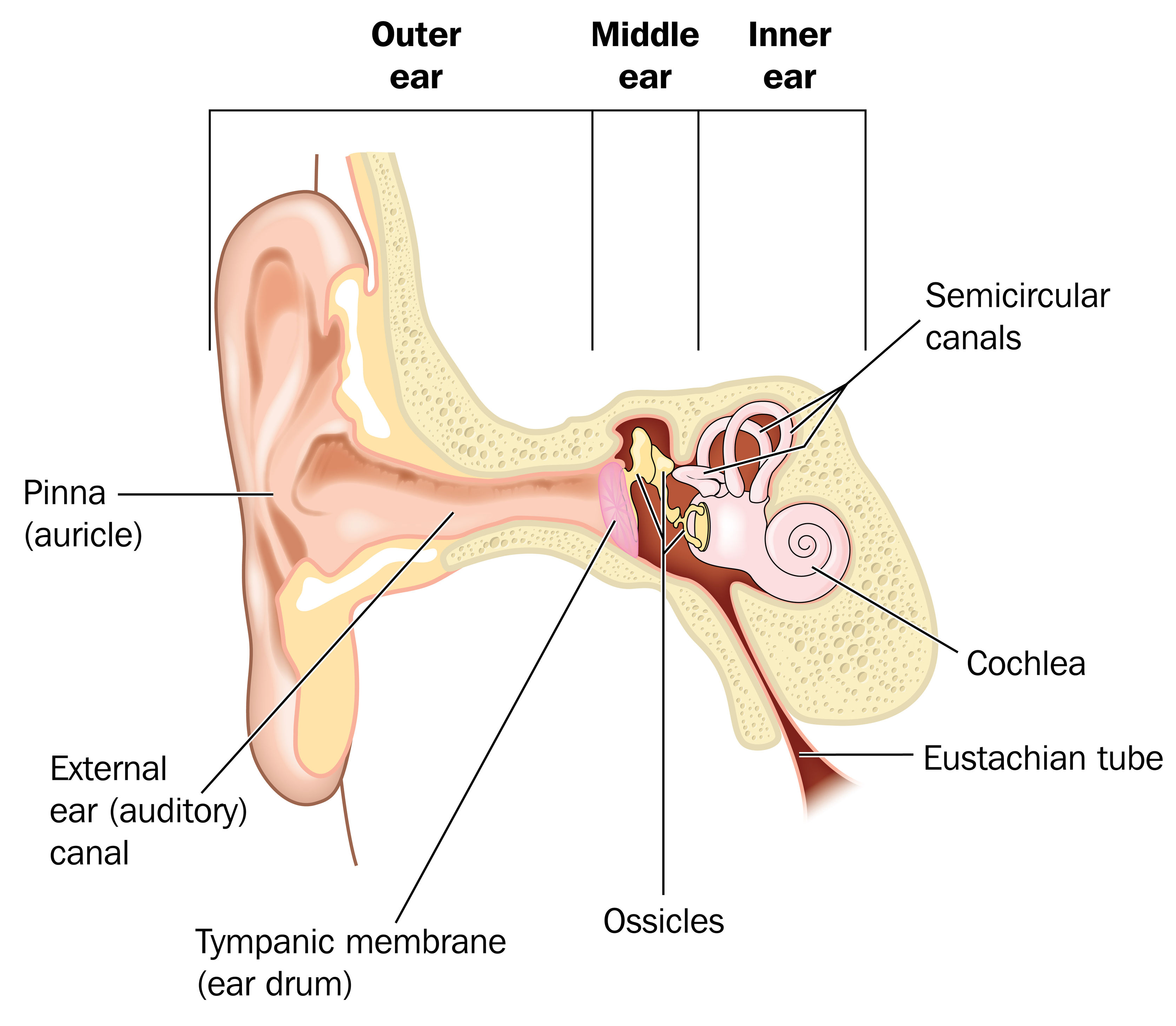
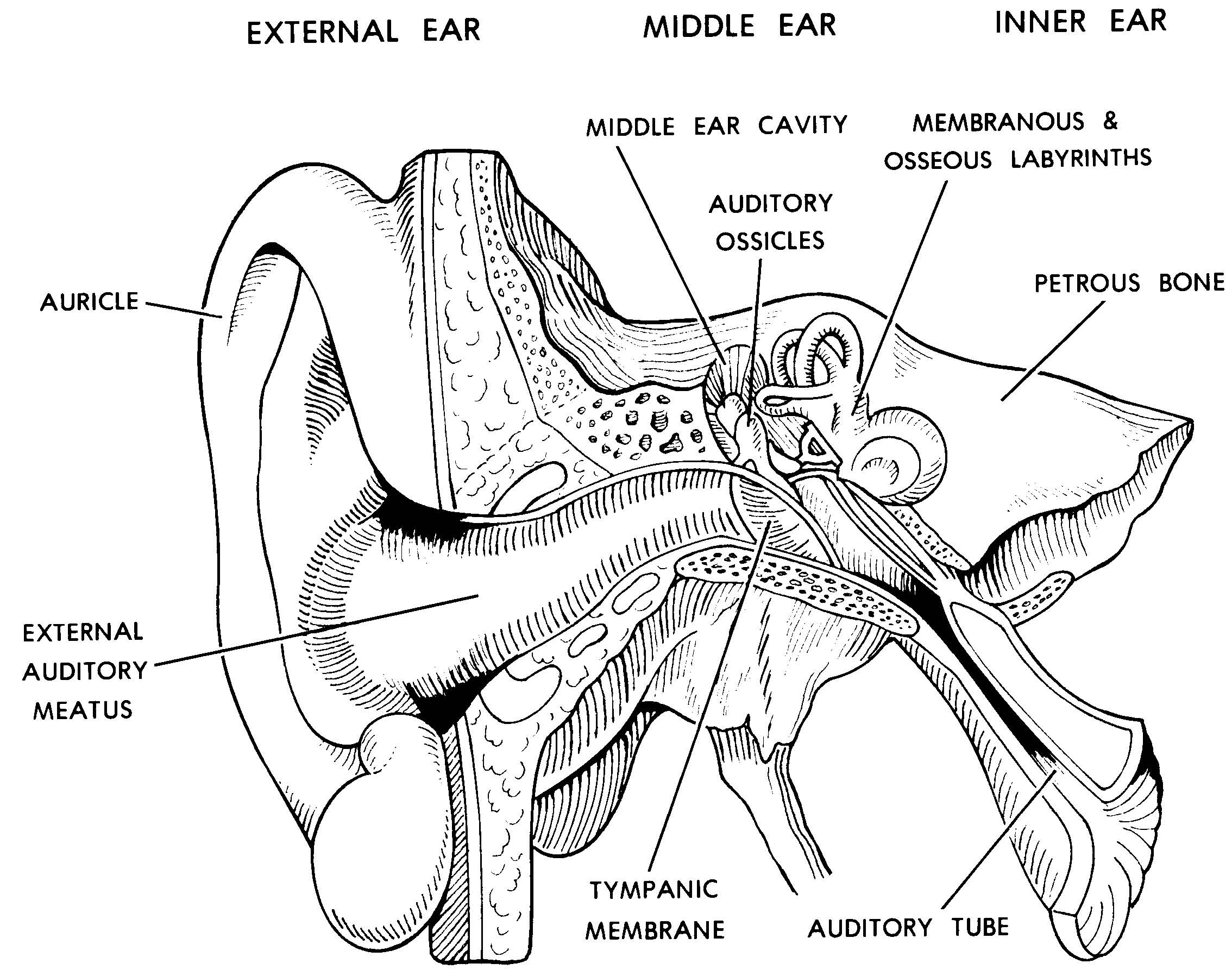
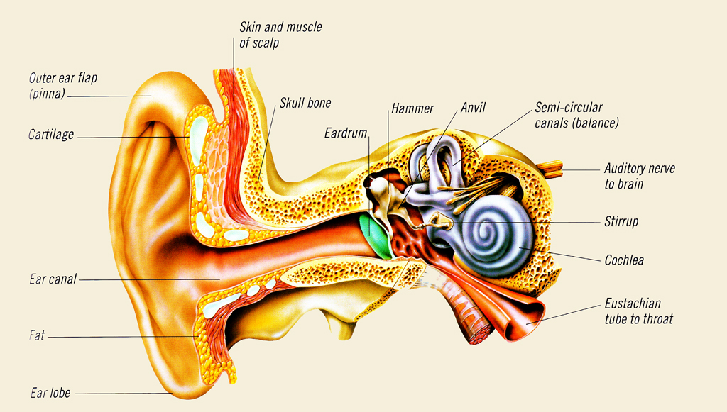
Ear diagrams (labeled and unlabeled) Overview image showing the structures of the outer ear and auditory tube Take a moment to look at the ear model labeled above. This shows you all of the structures you've just learned about in the video, labeled on one diagram.
Understanding how the ear works Hearing Link Services
Your inner ear is the last stop that sound waves make in a carefully orchestrated journey that starts from your outer ear. These waves travel from your outer ear through your middle ear to your inner ear. In the inner ear, the sound waves are converted into electrical energy, which your hearing nerve delivers to your brain as sound, making it.
Ear infections explained Dr Mark McGrath
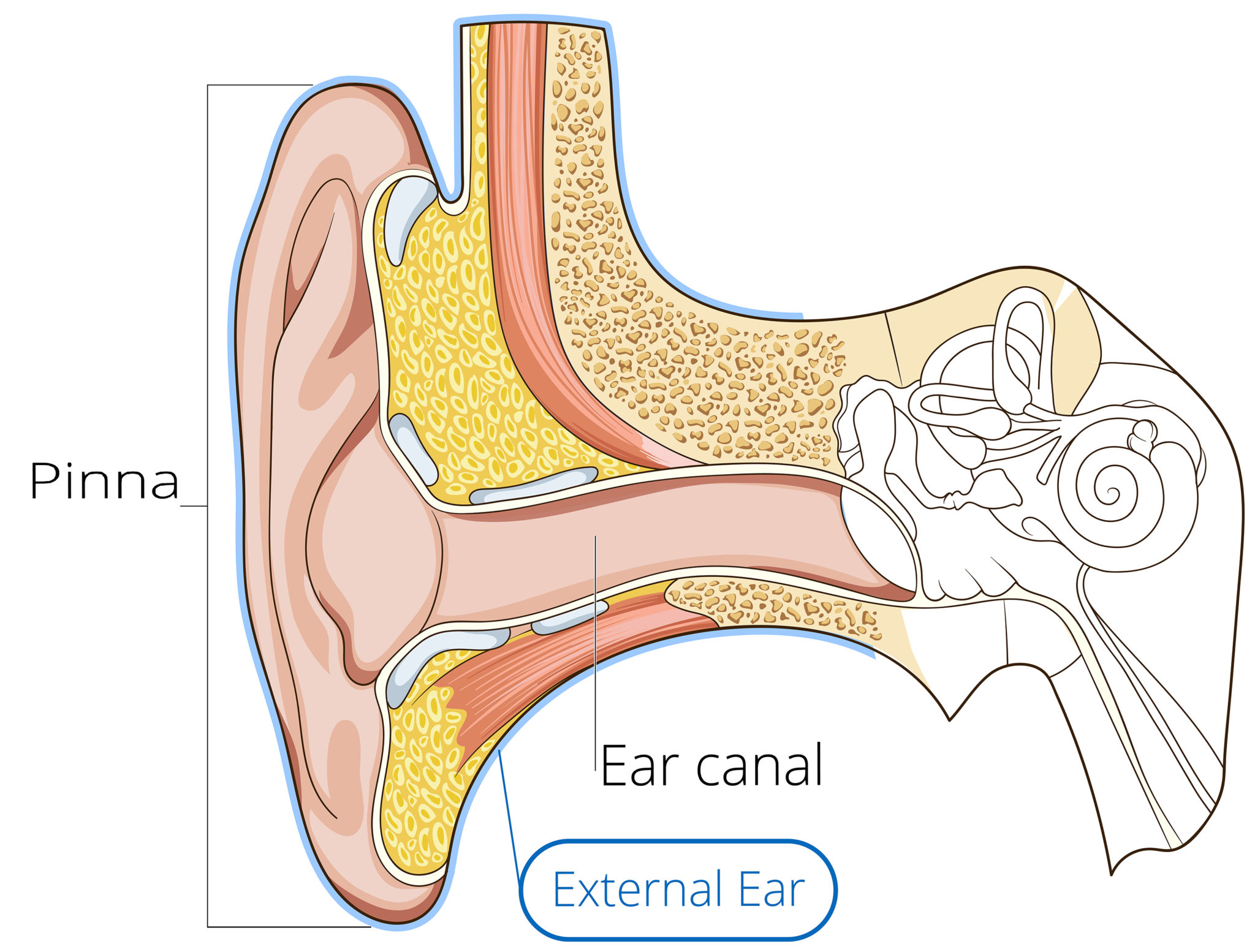
EAR CANAL The ear canal starts at the outer ear and ends at the ear drum. The canal is approximately an inch in length. The skin of the ear canal is very sensitive to pain and pressure. Under the skin the outer one third of the canal is cartilage and inner two thirds is bone. EAR DRUM
Images 11. Nervous System Basic Human Anatomy
labeling the ear by nielsejo86 185,927 plays 12 questions ~30 sec English 12p More 119 3.89 (you: not rated) Tries Unlimited [?] Last Played December 4, 2023 - 03:07 am There is a printable worksheet available for download here so you can take the quiz with pen and paper. Remaining 0 Correct 0 Wrong 0 Press play! 0% 0:00.0 Other Games of Interest
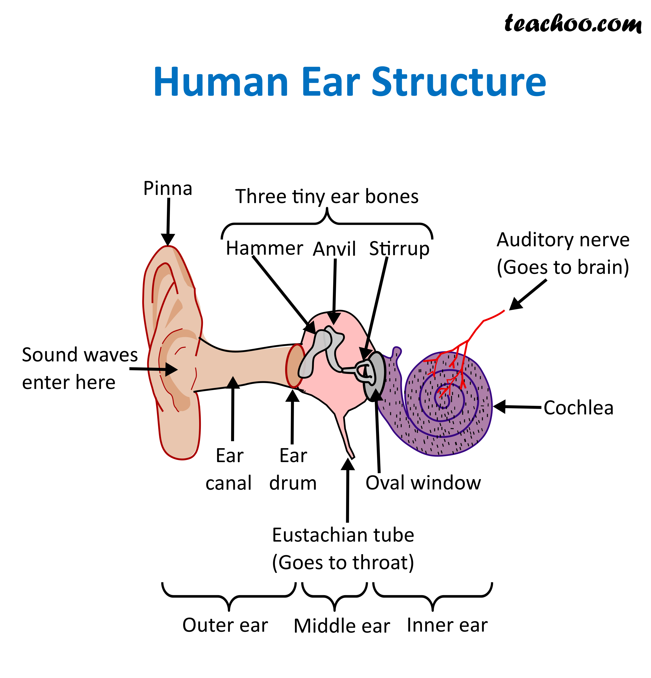
Structure and Function of Human Ear with Diagram Teachoo
Helix: The outermost curvature of the ear, extending from where the ear joins the head at the top to where it meets the lobule. The helix begins the funneling of sound waves into the ear; Fossa, superior crus, inferior crus, and antihelix: These sections make up the middle ridges and depressions of the outer ear. The superior crus is the first ridge that emerges moving in from the helix.
How You Hear Northland Audiology
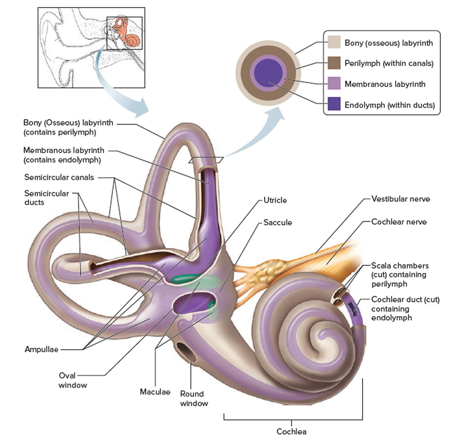
Anatomy Structure The ear is made up of the outer ear, middle ear, and inner ear. The inner ear consists of the bony labyrinth and membranous labyrinth. The bony labyrinth comprises three components: Cochlea: The cochlea is made of a hollow bone shaped like a snail and divided into two chambers by a membrane.

The Anatomy of the Outer Ear Health Life Media
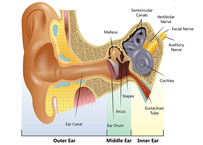
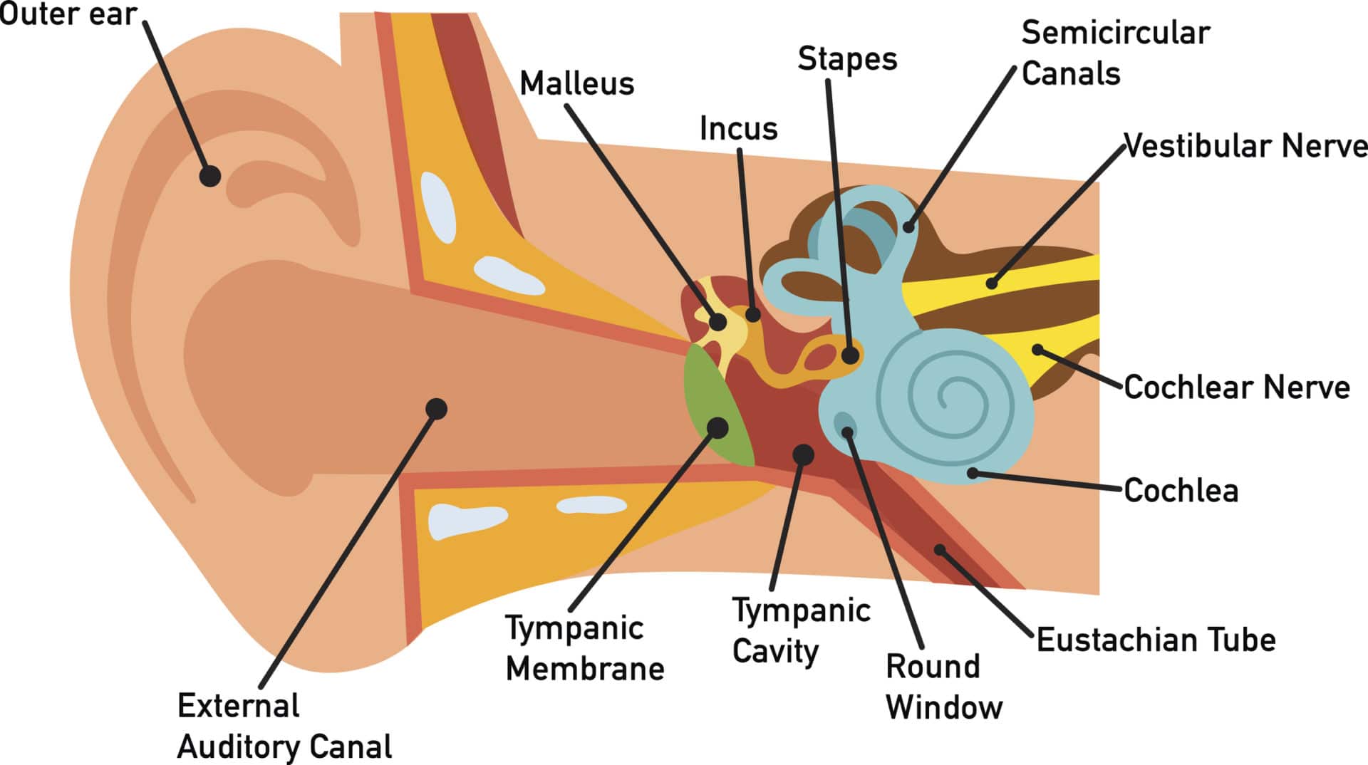
The inner ear is the innermost part of the ear and consists of the cochlea, auditory nerve, vestibule and semicircular canals. The inner ear is a maze of tubes and passages, referred to as the labyrinth. The inner ear is mainly responsible for balance and detecting sound. The cochlea contains the cells responsible for hearing, the auditory.
Your Hearing Heritage Hearing
A brief description of the human ear along with a well-labelled diagram is given below for reference. Well-Labelled Diagram of Ear The External ear or the outer ear consists of Pinna/auricle is the outermost section of the ear. The external auditory canal links the exterior ear to the inner or the middle ear.
How We Hear Hearing Associates, Inc.
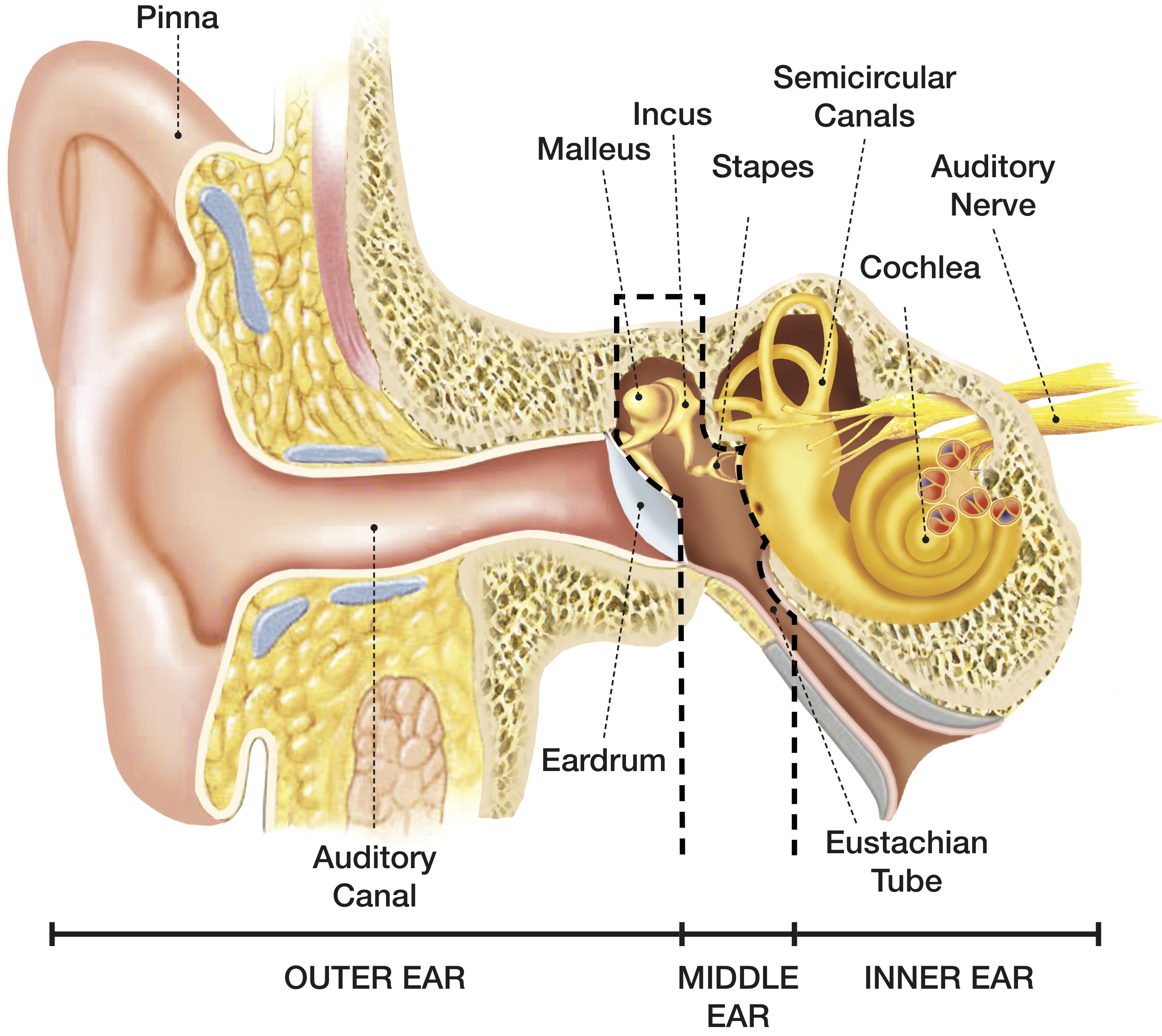
The parts of the ear include: External or outer ear, consisting of: Pinna or auricle. This is the outside part of the ear. External auditory canal or tube. This is the tube that connects the outer ear to the inside or middle ear. Tympanic membrane (eardrum). The tympanic membrane divides the external ear from the middle ear.

Hearing and the Structure of the Ear
The middle ear includes the eardrum, malleus, incus, and stapes. The inner ear includes semicircular canals, eustachian tube, cochlea, and vestibule and auditory nerves.">.

15.3 Hearing Anatomy & Physiology
The outer ear consists of the visible portion called the auricle, or pinna, which projects from the side of the head, and the short external auditory canal, the inner end of which is closed by the tympanic membrane, commonly called the eardrum. The function of the outer ear is to collect sound waves and guide them to the tympanic membrane.
Discovering Something New ongoing learning How the ear works
Meatus acusticus externus 1/4 Synonyms: External auditory meatus, External acoustic pore , show more. The ear is a complex part of an even more complex sensory system. It is situated bilaterally on the human skull, at the same level as the nose. The main functions of the ear are, of course, hearing, as well as constantly maintaining balance.
Human Ear Anatomy Parts of Ear Structure, Diagram and Ear Problems
Here is a blank human ear diagram for you to label, so that you can memorize the different parts of this vitally necessary organ, for good.
Ear Anatomy Causes of Hearing Loss Hearing Aids Audiology
Overview Your outer ear and middle ear are separated by your eardrum, and your inner ear houses the cochlea, vestibular nerve and semicircular canals (fluid-filled spaces involved in balance and hearing). What is the ear? Your ears are organs that detect and analyze sound. Located on each side of your head, they help with hearing and balance.